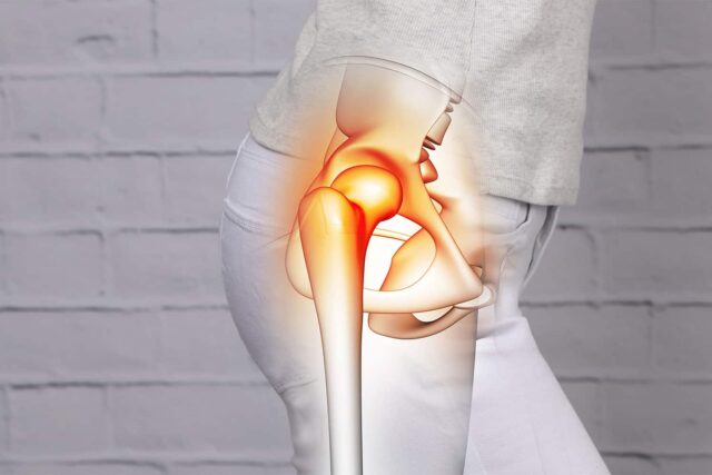
The main problem in this condition is the degeneration of the cartilage covering the ends of the bones. The loss of cartilage and its shock absorbing, cushioning effect leads to the bones coming closer to each other and their thickening in an attempt to protect the joint. Bony growth called osteophytes form around the joint edges. The joints and the surrounding ligaments, tissues may also develop swelling (inflammation).
Pain in arthritis may result from
Inflammation (swelling) of joints
Damage of joint tissues
Overloading of surrounding structures such as the muscles and ligaments
fatigue and extra effort required in mobilisation
Many people are not able to able to identify the symptoms of hip arthritis and often ignore it as a minor injury which would heal in a few days. They are unable to appreciate that the arthritis pain is not the same and cannot be treated like any other injury pain. Some of the other causes of hip pain include
Rheumatoid arthritis. This is an inflammatory condition which can involve the hip join, although is less common compared to OA. It is seen more in middle aged or elderly women.
Avascular necrosis (AVN) – In this condition the bone forming head of the thigh bone (femur) dies leading to bone collapse and severe hip damage.
What all are at risk of developing hip arthritis?
There are several factors that contribute to one’s chances of is developing this condition. These can be classified as those related to an individual and those related to the joint.
Individual factors include
Age over 50 years
Being overweight
Gender. Below the age of 50 years hip OA is more prevalence in men in, whereas above the age of 50 years it is more prevalent in women
Genetic predisposition
Occupation. Certain occupations such as farming which involve hard repetitive tasks, heavy lifting or standing for long periods are more at risk
Joint related factors such as
Joint shape. Abnormal shape predisposes the joint to more stress
Previous injury or surgery of the hip
Illness and infection – joint infection and conditions such as gout can contribute to the development of arthritis
Muscle function
What are the symptoms of hip arthritis?
Common symptoms include
Pain. This may be felt over the hip, groin or buttock and is described as a poorly localised, dull, aching sensation. It can radiate along the thigh towards the knee. Initially the pain is present only on movement or when the joint is loaded. As the condition progresses pain becomes more frequent and persists even during periods of rest. Some individuals find their arthritis pain increases in the winter months and rainy season.
Stiffness. This is worse in the morning and after periods of rest.
Reduced range of hip movements can make simple tasks such as walking or bending a challenge. Hip movements may be accompanied by a creaking or clicking noise/ sensation.
How is hip arthritis diagnosed?
Your doctor can often diagnose osteoarthritis on the basis of your symptoms and examination findings. Tests such as x-rays are commonly requested to confirm the diagnosis. Other tests such as blood tests, CT scan or MRI may also be requested for further evaluation.
What are the non-surgical pain relief options in hip arthritis?
The treatment of hip OA begins with education and self-management. The self-management options for hip arthritis include:
Weight loss
Balancing rest with activities (pacing) and avoiding high impact activities. Sitting crossed leg or on low height chairs should also be avoided if it increases your pain
Use of hot/ cold compression
Staying active. Regular gentle exercises such as walking, yoga, pilates, swimming can improve function, reduce pain, protect the joints and maintain muscle strength. Inactivity can worsen the arthritis symptoms
Wearing supportive footwear to cushion the impact on the hip joint and use of aids such as a walking stick
Your pain specialist can help with
Medications such as anti-inflammatories. These should be used medical supervision in the lowest dose and for the shortest duration possible, as long-term use is associated with risks. There are other painkillers which work through different mechanisms and can be prescribed by your pain specialist.
Joint injections
Steroid injections are the most commonly used injection option. As hip joint is a deep joint it is recommended that these injections are performed using ultrasound or x-ray guidance. Ultrasound guidance is preferred as it can be easily performed in the clinic and does not involve radiation exposure. Ultrasound helps to improve accuracy and reduce chances of complications. Most often a mixture of local anaesthetic and steroid is used for the injection. Local anaesthetics help to relax the muscle and steroids aid in reducing inflammation thus prolonging the effect of the injection. These injections also have a diagnostic role in confirming the source of pain.
PRP injections. The use of platelet-rich plasma for hip OA is under investigation in clinical trials with limited guidance available on their clinical role.
Treatments that are NOT recommended include
Supplements such as Glucosamine/Chondroitin, vitamin D and antioxidants are not recommended for hip arthritis
Hyaluronic acid injections. There is no strong to support their use. As per current evidence these injections are no better that placebo in improving function, stiffness and pain in hip OA.
What are the latest treatment options for hip joint pain?
When it comes to managing hip pain, there is a large lacuna in the treatment options as medications often offer limited relief or are limited by side effects and surgery is not always the answer as many are unwilling or unfit to have one. Some in their 40s and 50s are considered to be too young to undergo a joint replacement. Besides 5 to 15% of patients have persisting pain even after having a hip replacement.
For all these subgroups the new options including radiofrequency ablation, cooled radiofrequency and cryoablation offer a ray of hope. These options have been used most often for hip arthritis pain (as it is more common) although they work equally well for other hip joint pain conditions such as avascular necrosis (AVN).
The primary aim in these treatments remains the interruption or reduction of the pain signals travelling from the hip joint to the brain. This is achieved by deactivating or stunning the nerves responsible for transmitting the pain signals from these joints.
The method used for achieving deactivation is where these treatments differ. These options can be considered for anyone with hip joint pain who has not responded well to non-surgical or surgical options.
Typical indications include someone who Is not fit for hip replacement Doesn’t want to have surgery or is old and doesn’t want to go through extensive recovery and rehabilitation
Is young and wants to delay the surgery as worries about wearing out of hip joint and need for repeat surgery are not unfounded
Has persisting pain after hip replacement surgery
Has chronic hip pain with inadequate response to other treatment options
All the above mentioned treatment options share some common advantages such as
Minimally invasive pain management alternative for those not keen on surgery
Safe procedures without the increased risk of complications Can provide quick, lasting pain relief.
Less pain can translate into improve functional ability and reduced painkiller requirements, disability
Day care procedure with no requirement for overnight hospital stay
No cuts or incisions involved in the procedure
No requirement for general anaesthesia
Quick return to routine activities with no downtime and no requirement of prolonged rehabilitation
Diagnostic test injections are often performed prior to the radiofrequency procedure. This involves using small amount of local anaesthetic to numb the nerves in order to gauge the chances of success of the subsequent treatment. 50% or more reduction in pain after the test injection is considered a positive response and an indication to proceed with the treatment.
These new treatment options are discussed further in the section below
Radiofrequency ablation (RFA)
This procedure uses heat generated by radio waves to target specific nerves carrying pain from the hip joint. Specialised needles are used for controlled, targeted application of heat to these nerves, impairing their ability to send pain signals from hip to brain thereby resulting in pain relief.
The procedure involves placement of needles at specific locations under x ray and ultrasound guidance. For hip pain, branches of two specific nerves are targeted. These nerves are very small to be actually seen under ultrasound but are located in a specific area which is targeted using x-ray and ultrasound guidance.
Once the needles are correctly placed they are connected to the radiofrequency machine. As a safety precaution testing is done to ensure there is no other nerve close to the needle location. This is followed by the actual treatment involving the application of heat generated by radio waves.
Post procedure, pain relief usually begins within one to two weeks and may last up to 24 months. Like most medical treatments, individual responses to RFA treatment will vary with different levels of pain reduction. Those with a successful test injection have more chances of responding to the RFA treatment. Over time the nerves regenerate, at which point the treatment can be repeated if required. RFA is safe to administer repeatedly in patients with consistent pain reduction.
Cooled radiofrequency ablation (c-RFA)
This procedure involves placement of needles at the same location as described previously in the radiofrequency procedure. However the needles and the radiofrequency equipment used is different. Cooled Radiofrequency has water circulating through the device and the needle tip. cRFA is able to deliver more energy to the surrounding tissues, thereby creating a larger treatment area and increasing the chances of achieving effective pain relief.
Cryoablation
Cryoablation refers to the technique of inactivation or blocking the nerves for a long time by freezing the nerves sending pain signals to the brain. The procedure is performed using a special probe called cryoprobe, which is a hollow needle through which the super-cooled gas such as nitrous oxide or carbon dioxide is delivered directly to the treatment area. The extremely low temperatures achieved in the area being treated can inactivate the nerves thereby reducing the pain.
The probe is guided to the correct location using ultrasound and x-rays. Prior to cryoablation the nerve is identified with the help of a stimulation tests. Gas is delivered once the probe is in the correct place. An ice ball is created by a rapid decrease in the temperature at the tip of the probe and this can be visualised using ultrasound. Once the procedure is finished, the cryoprobe is removed and the procedure site covered with a small bandage.


